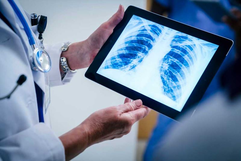
Chest X-Ray
Quick Facts
- A chest X-ray shows an image of the heart, lungs and chest bones.
- It might be ordered if you are having chest pain.

What is a chest X-ray?
A chest X-ray is a picture of the heart, lungs and chest bones. But it doesn’t show the inside structures of the heart.
Why is it done?
A chest X-ray shows the location, size and shape of the heart, lungs and some blood vessels. It is done when you have symptoms that may include:
- A persistent cough
- Chest pain
- Difficulty breathing
- Fever
- Coughing up blood
How is it done?
You will be positioned next to the X-ray film. You might wear a hospital gown over your chest. An X-ray machine is turned on for a fraction of a second. During this time, a small beam of X-rays passes through the chest and makes an image on special photographic film. Sometimes, two pictures are taken — a front and side view. Or, your cardiologist (heart doctor) might need additional views. X-ray results are often ready right away.
Does it hurt, or is it harmful?
No, you won’t feel the X-rays as the pictures are taken.
The amount of radiation used in a chest X-ray is small. It is about the same amount of radiation people are exposed to naturally over 10 days. This small amount of radiation isn’t considered dangerous. However, pregnant women should avoid even this low level of radiation when possible.
There are usually no restrictions on what you can do after an X-ray.


